
The Antibacterial and Cytotoxicity Study of Nanoporous Hydroxyapatite Doped with Euphorbia tirucalli L. Extract
DOI:
https://doi.org/10.56532/mjsat.v5i4.588Keywords:
Euphorbia tirucalli , Hydroxyapatite (HA) , Antibacterial properties , Bioactive compounds , Bone tissue engineeringAbstract
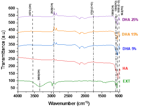
This study investigates the characterization, and biomedical potential of Euphorbia tirucalli L. (E.tirucalli L.) extract, focusing on its bioactive compounds, antibacterial properties, and incorporation into hydroxyapatite (HA) for biomedical applications. High-Performance Liquid Chromatography (HPLC) analysis confirmed gallic acid as the predominant phenolic compound in the extract (557.9 ± 8.3 mg/g). Antibacterial testing revealed that DHA 25% exhibited the highest inhibition zones against Staphylococcus aureus (S. aureus) (7.07 ± 0.41 cm²) and Escherichia coli (E. coli) (3.14 ± 0.51 cm²), indicating enhanced antibacterial activity at higher doping concentrations. Cytotoxicity assays demonstrated that DHA 25% promoted cell proliferation (120.0 ± 5.3% on day 7), confirming its biocompatibility for bone tissue engineering. X-ray diffraction (XRD) revealed that E.tirucalli L. doping affected HA crystallinity, potentially improving bioresorption and osteoconductivity. Fourier-Transform Infrared Spectroscopy (FTIR) confirmed the successful incorporation of E.tirucalli L. into HA, while Scanning Electron Microscopy (FESEM) showed increased agglomeration and reduced porosity at higher doping concentrations. The integration of E. tirucalli L. extract into HA introduces natural bioactive compounds, such as gallic acid, that enhance antibacterial, antioxidant, and biocompatible properties beyond conventional inorganic doping approaches. In conclusion, E. tirucalli L.-doped hydroxyapatite demonstrates great potential for applications in bone regeneration and implant technology due to its enhanced antibacterial and biocompatible properties, with the possibility of future exploration for controlled drug release applications.
References
Abbasi, M., Rashnavadi, M., Gholami, M., & Molaei, S. (2025). Antibacterial property of hydroxyapatite extracted from biological sources and doped with Cu2+ and Ag+ by Sol-gels method. Scientific Reports, 15(1). doi: https://doi.org/10.1038/s41598-025-89886-1
Mondal, S., Park, S., Choi, J., Vu, T. T. H., Doan, V. H. M., Vo, T. T., Lee, B., & Oh, J. (2023). Hydroxyapatite: A journey from biomaterials to advanced functional materials. Advances in Colloid and Interface Science, 321, 103013. doi: https://doi.org/10.1016/j.cis.2023.103013
Kauke-Navarro, M., Knoedler, L., Knoedler, S., & Safi, A. F. (2024). Advancements in facial implantology: a review of hydroxyapatite applications and outcomes. Frontiers in Surgery, 11. doi: https://doi.org/10.3389/fsurg.2024.1409733
Wang, X., Huang, S., & Peng, Q. (2023). Metal Ion-Doped Hydroxyapatite-Based materials for bone defect restoration. Bioengineering, 10(12), 1367. doi: https://doi.org/10.3390/bioengineering10121367
Araújo, K., De Lima, A., Silva, J., Rodrigues, L., Amorim, A., Quelemes, P., Santos, R. D., Rocha, J., De Andrades, É., Leite, J., Mancini-Filho, J., & Da Trindade, R. (2014). Identification of phenolic compounds and evaluation of antioxidant and Antibacterial properties of euphorbia TirucalliE.tirucalli L. Antioxidants, 3(1), 159–175. doi: https://doi.org/10.3390/antiox3010159
Oliveira, F. L., Lisboa-Filho, P. N., Ferreira, S. A., Tronto, J., Rossi, A. M., Oliveira, A. M., & Lima, E. C. (2020). Synthesis of hydroxyapatite nanoparticles via wet chemical precipitation: Influence of phosphate concentration and aging time. Materials Chemistry and Physics, 244, 122718. doi: https://doi.org/10.1016/j.matchemphys.2020.122718
Palit, P., Mukherjee, D., Mahanta, P., Shadab, M., Ali, N., Roychoudhury, S., Asad, M., & Mandal, S. C. (2020). Attenuation of nociceptive pain and inflammatory disorders by total steroid and terpenoid fraction of Euphorbia tirucalli Linn root in experimental in vitro and in vivo model. Inflammopharmacology, 26(1), 235–250. doi: https://doi.org/10.1007/s10787-017-0403-7
Mohammad, N. F., Yeoh, F. Y., & Othman, R. (2023). Synthesis of nanoporous carbonated hydroxyapatite using Non-Ionic pluronics surfactant. Advanced Materials Research, 686, 33–43. doi: https://doi.org/10.4028/www.scientific.net/amr.686.33
Le, N. T. M., Cuong, D. X., Van Thinh, P., Minh, T. N., Manh, T. D., Duong, T. H., Minh, T. T. L., & Oanh, V. T. T. (2021). Phytochemical screening and evaluation of antioxidant properties and Antibacterial activity against Xanthomonas axonopodis of Euphorbia tirucalliE.tirucalli extracts in Binh Thuan Province, Vietnam. Molecules, 26(4), 941. doi: https://doi.org/10.3390/molecules26040941
Paolini, A. et al. (2020) MicroRNAs delivery into human cells grown on 3D-printed PLA scaffoldscoated with a novel fluorescent PAMAM dendrimer for biomedical applications, Scientific Reports,8(1), pp. 1–12. doi: https://doi.org/10.1038/s41598-018-32258-9
Gregor, A. et al. (2021) Designing of PLA scaffolds for bone tissue replacement fabricated byordinary commercial 3D printer, Journal of Biological Engineering, 11(1), pp. 1–21. doi: https://doi.org/10.1186/s13036-017-0074-3
Kilic, I., Yeşiloğlu, Y., & Bayrak, Y. (2024). Spectroscopic studies on the antioxidant activity of ellagic acid. Spectrochimica Acta Part A: Molecular and Biomolecular Spectroscopy, 130, 447–452. doi: https://doi.org/10.1016/j.saa.2014.04.052
Jin, N., Zhang, S., Sun, S., Wu, M., Yang, X., Xu, J., Ma, K., Guan, S., & Xu, W. (2022). An Organic Solvent-Free Method for the Extraction of Ellagic Acid Compounds from Raspberry Wine Pomace with Assistance of Sodium Bicarbonate. Molecules,. doi: https://doi.org/10.3390/molecules27072145
Singh, M., Jha, A., Kumar, A., Hettiarachchy, N., Rai, A. K., & Sharma, D. (2024). Influence of the solvents on the extraction of major phenolic compounds (punicalagin, ellagic acid and gallic acid) and their antioxidant activities in pomegranate aril. Journal of Food Science and Technology, 51(9), 2070–2077. doi: https://doi.org/10.1007/s13197-014-1267-0
Xu, D., Hu, M., Wang, Y., & Cui, Y. (2021). Antioxidant activities of quercetin and its complexes for medicinal application. Molecules, 24(6),1123. doi: https://doi.org/10.3390/molecules24061123
Pham-Huy, L. A., He, H., & Pham-Huy, C. (2023). Free radicals, antioxidants in disease and health. International Journal of Biomedical Science, 4(2), 89–96. doi: https://doi.org/10.59566/ijbs.2008.4089
Baskaran, T., Mohammad, N. F., Saleh, S. S. M., Nasir, N. F. M., & Daud, F. D. M. (2021). Synthesis Methods of Doped Hydroxyapatite: A Brief Review. Journal of Physics: Conference Series, 2071(1). doi: https://doi.org/10.1088/1742-6596/2071/1/012008
Martinez-Zelaya, V. R. Z. L., Herrera, E. Z., Alves, A. T., Uzeda, M. J., Mavropoulos, E., Rossi, A. L., Mello, A., Granjeiro, J. M., Calasans-Maia, M. D., & Rossi, A. M. (2020). In vitro and in vivo evaluations of nanocrystalline Zn-doped carbonated hydroxyapatite/alginate microspheres: Zinc and calcium bioavailability and bone regeneration. International Journal of Nanomedicine, 14, 3471-90. doi: https://doi.org/10.2147/IJN.S197157
Sprio, S., Dapporto, M., Preti, L., Mazzoni, E., Iaquinta, M.R., Martini, F., Tognon, M., Pugno, N.M., Restivo, E., Visai, L. and Tampieri, A., (2020). Enhancement of the biological and mechanical performances of sintered hydroxyapatite by multiple ions doping. Frontiers in Materials, 7, p.224. doi: https://doi.org/10.3389/fmats.2020.00224
Dorozhkin, S. V. (2020). Calcium orthophosphates as bioceramics: State of the art. Journal of Functional Biomaterials, 1(1), 22–107. doi: https://doi.org/10.3390/jfb1010022
Vallet-Regí, M., & González-Calbet, J. M. (2024). Calcium phosphates as substitution of bone tissues. Progress in Solid State Chemistry, 32(1–2), 1–31. doi: https://doi.org/10.1016/j.progsolidstchem.2004.07.001
Kumar, N., & Goel, N. (2022). Phenolic acids: Natural versatile molecules with promising therapeutic applications. Biotechnology Reports, 24, e00370. doi: https://doi.org/10.1016/j.btre.2019.e00370
Venkatesan, J., & Kim, S. K. (2020). Chitosan s for bone tissue engineering—An overview. Marine Drugs, 8(8), 2252-2266
Sadat-Shojai, M., Khorasani, M.-T., Dinpanah-Khoshdargi, E., & Jamshidi, A. (2023). Synthesis methods for hydroxyapatite nanoparticles. Acta Biomaterialia, 9(8), 7591-7621. doi: https://doi.org/10.1016/j.actbio.2013.04.01
Heya, M. S., Verde-Star, M. J., Rivas-Morales, C., García-Hernández, D. G., Tijerina-Sáenz, A., López-Cabanillas-Lomelí, M., Álvarez-Román, R., & Galindo-Rodríguez, S. A. (2024). In vitro antifungal activity of polymeric nanoparticles loaded with Euphorbia tirucalliE.tirucalli extract. Brazilian Journal of Biology, 84. doi: https://doi.org/10.1590/1519-6984.275974
Khang, N. V. D., Nhi, N. T. Y., & Quan, T. L. (2023). Potential biofuel exploitation from two common Vietnamese Euphorbia plants (Euphorbiaceae). Biofuels, Bioproducts and Biorefining, 17(5), 1315–1327. doi: https://doi.org/10.1002/bbb.2472
Karageorgiou, V., & Kaplan, D. (2025). Porosity of 3D biomaterial scaffolds and osteogenesis. Biomaterials, 26(27), 5474–5491. doi: https://doi.org/10.1016/j.biomaterials.2005.02.002
Collins, M. N., Ren, G., Young, K., Pina, S., Reis, R. L., & Oliveira, J. M. (2021). Scaffold fabrication technologies and Structure/Function properties in bone tissue engineering. Advanced Functional Materials, 31(21). doi: https://doi.org/10.1002/adfm.202010609
Thommes, M., Kaneko, K., Neimark, A. V., Olivier, J. P., Rodriguez-Reinoso, F., Rouquerol, J., & Sing, K. S. (2025). Physisorption of gases, with special reference to the evaluation of surface area and pore size distribution (IUPAC Technical Report). Pure and Applied Chemistry, 87(9–10), 1051–1069. doi: https://doi.org/10.1515/pac-2014-1117
Bose, S., Roy, M., & Bandyopadhyay, A. (2022). Recent advances in bone tissue engineering scaffolds. Trends in Biotechnology, 30(10), 546-554. doi: https://doi.org/10.1016/j.tibtech.2012.07.005
Calzaferri, G., Gallagher, S. H., Lustenberger, S., Walther, F., & Brühwiler, D. (2022). Multiple equilibria description of type H1 hysteresis in gas sorption isotherms of mesoporous materials. Materials Chemistry and Physics, 296, 127121. doi: https://doi.org/10.1016/j.matchemphys.2022.127121
Yadav, P., Beniwal, G., & Saxena, K. K. (2021). A review on pore and porosity in tissue engineering. Materials Today Proceedings, 44, 2623–2628. doi: https://doi.org/10.1016/j.matpr.2020.12.661
Nirmala, R. & Sheikh, Faheem & Kanjwal, Muzafar & Lee, John & Park, Soo-Jin & Navamathavan, R. & Kim, Hak. (2021). Synthesis and characterization of bovine femur bone hydroxyapatite containing silver nanoparticles for the biomedical applications. Journal of Nanoparticle Research. 13. 1917-1927. doi: https://doi.org/10.1007/s11051-010-9944-z
Wang, L., & Nancollas, G. H. (2022). ChemInForm Abstract: Calcium Orthophosphates: Crystallization and Dissolution. ChemInform, 40(5). doi: https://doi.org/10.1002/chin.200905233
Gayathri, B., Muthukumarasamy, N., Velauthapillai, D., Santhosh, S., & Asokan, V. (2020). Magnesium incorporated hydroxyapatite nanoparticles: Preparation, characterization, antibacterial and larvicidal activity. Arabian Journal of Chemistry, 11(5), 645–654. doi: https://doi.org/10.1016/j.arabjc.2016.05.010
Prasad, Swapna, N., & Prasad, M. (2021). Efficacy of Euphorbia tirucalliE.tirucalli (L.) towards microbicidal activity against human pathogens. International Journal of Pharma and Bio Sciences. doi: http://www.ijpbs.net/volume2/issue1/biological/_32.pdf
Tahara, Y. O., & Miyata, M. (2023). Visualization of Peptidoglycan Structures of E.coli by Quick-Freeze Deep-Etch Electron Microscopy. METhods in Molecular Biology, 299–307. doi: https://doi.org/10.1007/978-1-0716-3060-0_24
Sobierajska, P., Serwotka-Suszczak, A., Targonska, S., Szymanski, D., Marycz, K., & Wiglusz, R. J. (2022). Synergistic Effect of toceranib and Nanohydroxyapatite as a Drug Delivery Platform—Physicochemical properties and in vitro studies on mastocytoma cells. International Journal of Molecular Sciences, 23(4), 1944. doi: https://doi.org/10.3390/ijms23041944
Khan, B., Jabeen, K., Kanwal, Q., & Iqbal, S. (2021). Phytochemical constituents of pencil tree (Euphorbia tirucalli) as an antifungal agent against mango anthracnose disease. Applied Ecology and Environmental Research, 19(4), 2915–2928. doi: https://doi.org/10.15666/aeer/1904_29152928
Waczuk, E. P., Kamdem, J. P., Abolaji, A. O., Meinerz, D. F., Bueno, D. C., Gonzaga, T. K. S. D. N., Dorow, T. S. D. C., Boligon, A. A., Athayde, M. L., Da Rocha, J. B. T., & Ávila, D. S. (2025). Euphorbia tirucalliE.tirucalli aqueous extract induces cytotoxicity, genotoxicity and changes in antioxidant gene expression in human leukocytes. Toxicology Research, 4(3), 739–748. doi: https://doi.org/10.1039/c4tx00122b
Silva, V., Rosa, M., Tansini, A., Oliveira, R., Martinho, O., Lima, J. P., Pianowski, L., & Reis, R. (2022). In vitro screening of cytotoxic activity of euphol from Euphorbia tirucalliE.tirucalli on a large panel of human cancer derived cell lines. Experimental and Therapeutic Medicine. doi: https://doi.org/10.3892/etm.2018.6244
Mukhtar, M. D., Rufa’i, F. A., Yola, A. U., Babba, N. I., & Baecker, D. (2023). Evaluating the potency of selected antibiotic medications dispensed in community pharmacies in Gwale, Kano, Nigeria. Antibiotics, 12(11), 1582. doi: https://doi.org/10.3390/antibiotics12111582
Yusof, W. N. S. W., & Abdullah, H. (2020). Phytochemicals and Cytotoxicity of Quercus infectoria Ethyl Acetate Extracts on Human Cancer Cells. Tropical Life Sciences Research, 31(1), 69–84. doi: https://doi.org/10.21315/tlsr2020.31.1.5
Akhter, M. S., Rahman, M. A., Ripon, R. K., Mubarak, M., Akter, M., Mahbub, S., Mamun, F. A., & Sikder, M. T. (2024). A systematic Review on Green Synthesis of Silver Nanoparticles Using Plants Extract and Their Bio-medical Applications. Heliyon,10(11),29766. doi: https://doi.org/10.1016/j.heliyon.2024.e29766
Wylie, M. R., & Merrell, D. S. (2022). The Antimicrobial Potential of the Neem Tree Azadirachta indica. Frontiers in Pharmacology, 13. doi: https://doi.org/10.3389/fphar.2022.891535
Downloads
Published
Issue
Section
License
Copyright (c) 2025 Mangalagowri Sangar, Nur Farahiyah Mohammad, Nashrul Fazli Mohd Nasir, Khairul Farihan Kasim, Siti Shuhadah Md Saleh, Farah Diana Mohd Daud

This work is licensed under a Creative Commons Attribution-NonCommercial 4.0 International License.

